Figure 1 from Brain surface temperature under a craniotomy.
Por um escritor misterioso
Descrição
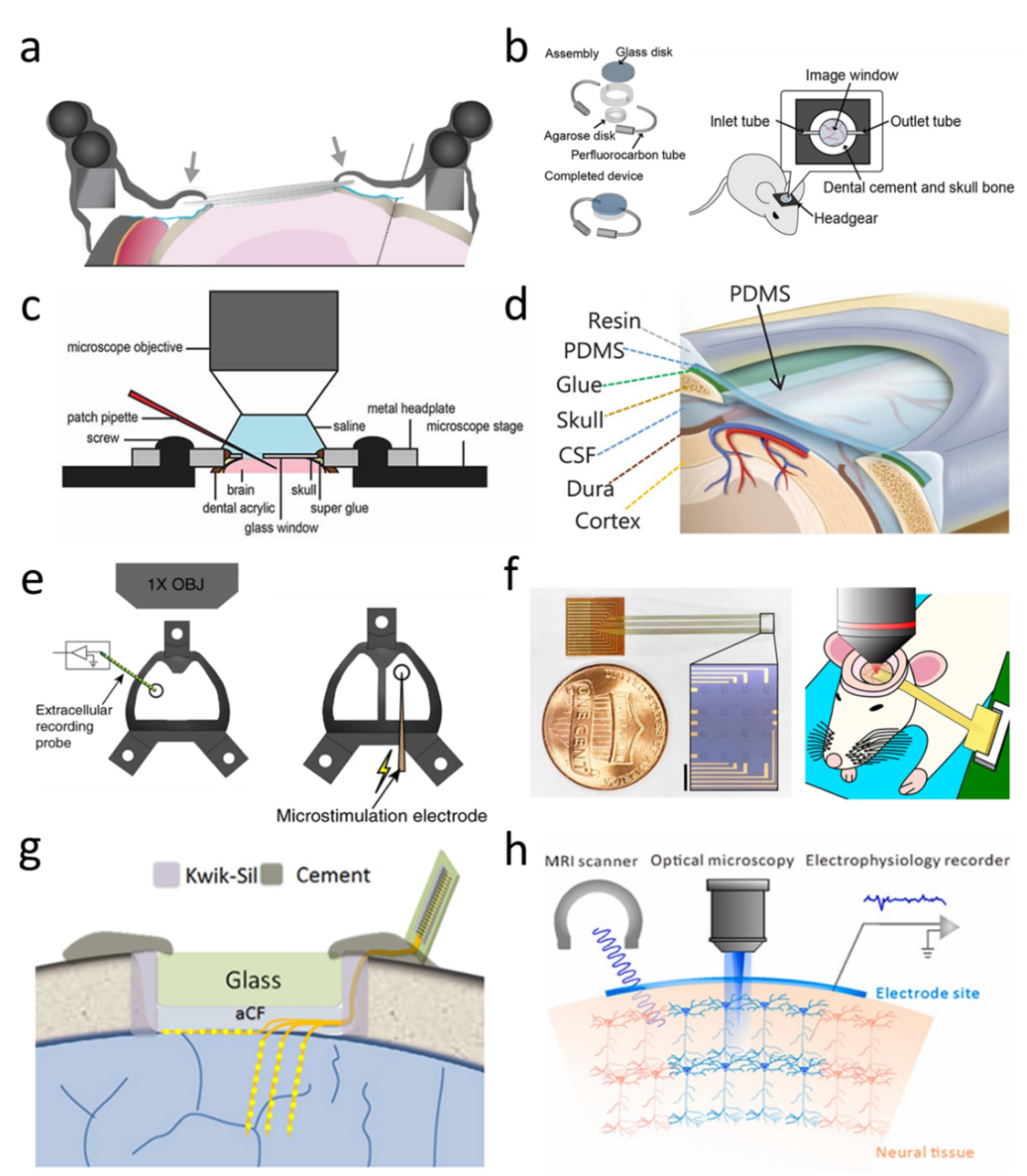
Fig. 1. Rapid cooling of the brain surface in an in vivo mouse preparation. A: schematic representation of a cranial window during recording of temperature and single-cell activity in the anesthetized mouse. The main potential routes of heat transfer are indicated. B: brain surface temperature measured with the thermocouple during replacement of the artificial cerebrospinal fluid (ACSF) with fresh ACSF warmed to 38°C. ACSF was replaced twice, indicated by the arrowheads. - "Brain surface temperature under a craniotomy."

Full article: Brain temperature and its role in physiology and pathophysiology: Lessons from 20 years of thermorecording
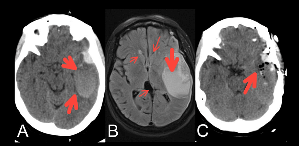
Cureus, Spontaneous Intracranial Hemorrhage Concurrent With Subarachnoid and Subdural Hemorrhages: Report of a Rare Case
Electronics, Free Full-Text

Applications of flexible electronics related to cardiocerebral vascular system - ScienceDirect
Brain Sciences, Free Full-Text

Regional temperature and quantitative cerebral blood flow responses to cortical spreading depolarization in the rat - Chunyan Li, Raj K Narayan, Ping Wang, Jed A Hartings, 2017

Thermal map of the brain. The results of measurements of the
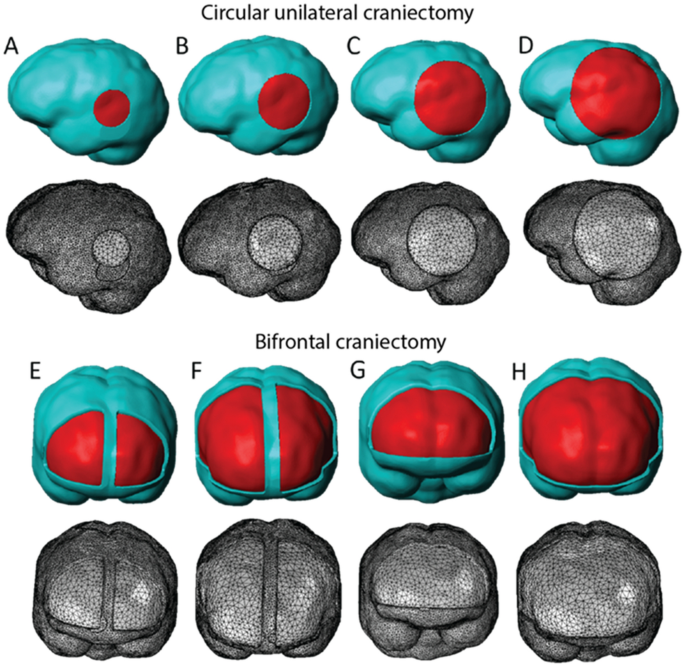
Decompressive craniectomy of post-traumatic brain injury: an in silico modelling approach for intracranial hypertension management
JCM, Free Full-Text

Temporal/Subtemporal Craniotomy

Correlation of core temperature and brain state. A: raw neocortical

Infrared thermography display of cortical temperature in cats

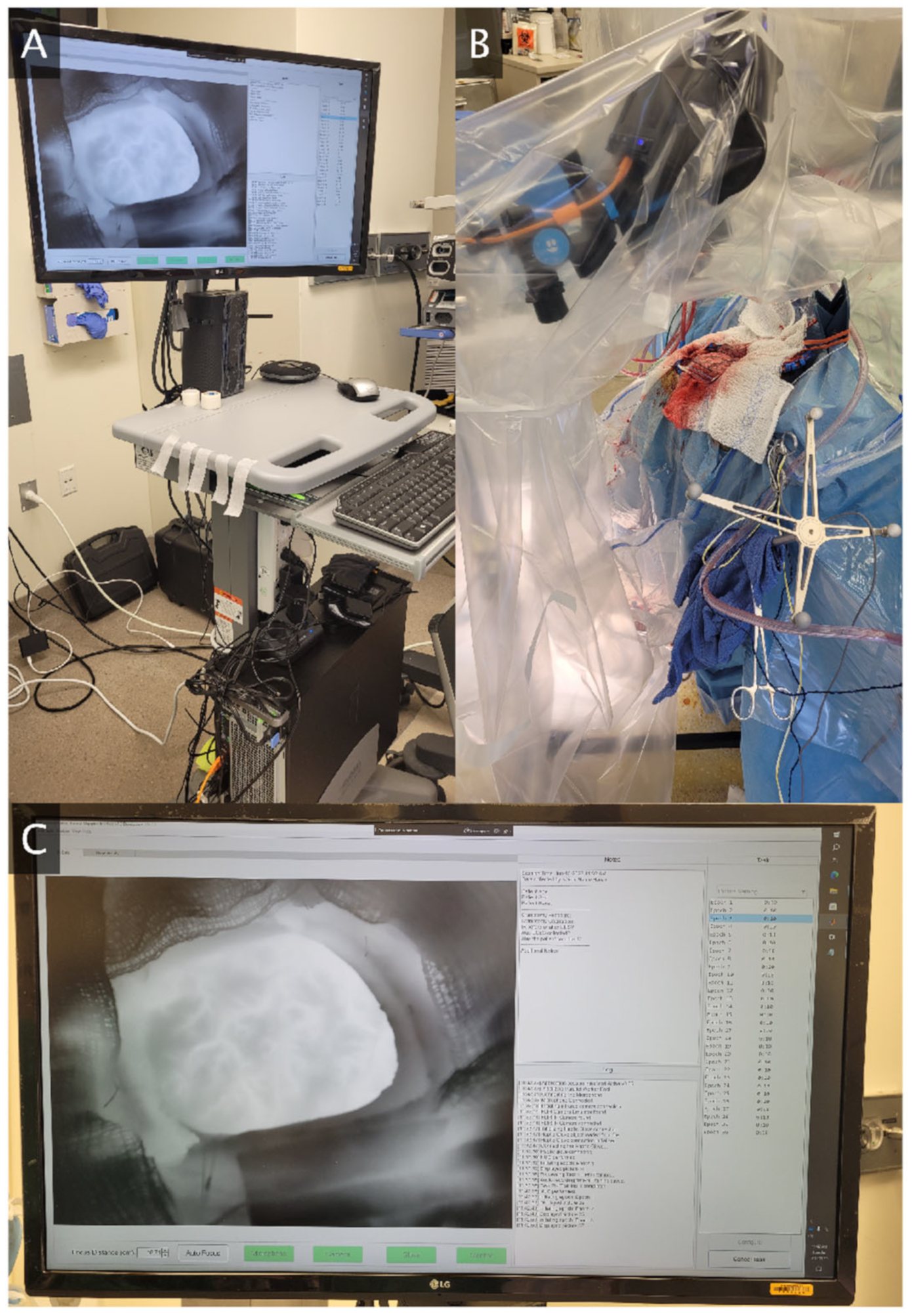
Data collection and craniotomy. Left: The infrared camera setup is
de
por adulto (o preço varia de acordo com o tamanho do grupo)